Equipment and services -Cellular and molecular imaging core facility

Location: Kunnskapsenter 3rd. floor, room 423.03.031 Confocal Imaging lab

This is a confocal laser scanning microscope that works well for all standard confocal applications. The microscope is also equipped with an Airyscan detector for gentle super resolution imaging.
General description:
This confocal microscope is well suited for studies requiring subcellular resolution. It is simple to set up with z stacks, multiple positions, tilescans or timelapse imaging, and has well-plate navigation for 96 and 24 well plates. Spectral unmixing can be performed based on fluorescence emission spectra. The airyscan detector allows for gentle super resolution imaging, and can be used in FAST or super resolution mode. Being a point scanning microscope, the LSM 880 gives less background deeper into samples compared to a spinning disk microscope, and allows adjusting pixel size independent of objective magnification.
Technical details:
- General: Definite focus, incubation chamber with temperature and CO2 control.
- Detectors: 2 PMTs (blue- and red-shifted), a GaAsP PMT spectral detector (array of 32 GaAsP PMTs) and an Airyscan detector. Photometrix Prime 95B camera for widefield imaging (only available in Zen Blue, while confocal imaging is controlled through Zen Black)
- Objectives: 5x, 10x and 20x air objecties, 40x water immersion objective, 40x|1.4 and 63x|1.4 oil immersion objectives
- Laser lines: 405, 458, 488, 514, 561 and 633nm
Location: Kunnskapssenter 3rd. floor, room 423.03.031 Confocal Imaging lab

This is a confocal microscope equipped with a STED module for super resolution, and a FLIM module for fluorescence lifetime imaging. The advanced modules are further described under special applications.
General description:
This is a confocal laser scanning microscope which is well suited for studies requiring subcellular resolution. The microscope is equipped with a white light laser (WLL), which allows to freely choose excitation wavelength in steps of 1nm. This is advantageous for samples with fluorophores that have excitation outside of the standard laser lines, and for samples with many fluroescent labels (multiplexing). Thanks to the WLL, labels can be distinguished through spectral unmixing based on both excitation and emission spectra, called "Lambda Square mapping".
A "navigator" software module allows automation of image acquisition, and fast sample overview. Z stacks, tilescans, multiple positions and timelapses can be acquired.
Technical details:
- General: High speed resonant scanner (8 KHz) and SuperZ galvo stage for fast lateral and axial acquisition; Adaptive Focus Control (AFC); incubation chamber with temperature and CO2 control.
- Detectors: 2 HyD (high sensitivity, hybrid GaAsP photon counting detectors) and 2 PMTs for confocal imaging. Hamamatsu ORCA-Flash 4.0 camera for widefield imaging.
- Objectives: 10x|0.4 (air), 10x|0.4 (multi), 20x|0.75 (multi), 40x|1.3 (oil), 63x|1.4 (oil), 63x|1.3 (glyc), 100x|1.4 (oil).
- Confocal Laser lines: 405nm, WLL2 (white light laser) tunable from 470-670nm.
- STED depletion lasers: 592nm, 660nm, 775nm.
Location: Kunnskapsenter 3rd. floor, room 423.03.003 Confocal Imaging lab

This is a state of the art spinning disk confocal, offering confocal resolution at speeds 10-100 times faster than the classic point scanning confocals.
General description:
Spinning disk confocal microscope ideal for acquisitions of large regions of interest, or of many cells or events for statistical analysis. The microscope is set up for dualcam imaging, where one frame can be captured with two colors simultaneously, ideal for large screens or for live acquisitions with fast kinetics. The microscope is surrounded by a black incubator where temperature, CO2 and oxygen concentration can be controlled, allowing to simulate biological conditions (including hypoxia) during live imaging. The microscope is also equipped with an automatic water dispenser (for water immersion objectives), that facilitates live imaging of specimens in buffer over large regions or over long time periods. For thin samples that don't require confocal optical sectioning, the microscope can easily be used as a high-end widefield fluroescence microscope.
Technical details:
- General: z piezo with 600μm range for fast z movement; incubation chamber with liquid reservoir (for humidity), CO2 and O2 (through N2) control; Perfect Focus Sysmte (PFS)
- Objectives: 4x, 10x, 20x and 40x air objectives, 40x and 60x water immersion objectives, 100x oil objective and 100x silicone oil objective.
- Detectors: two Photometrics Kinetix back illuminated sCMOS cameras for single or dualcam fluorescenec detection. One high-end color camera for colored slides (e.g. H&E stained histology samples).
- Laser lines: 405nm, 446nm, 477nm, 518nm, 546nm, 638nm, 749nm
Location: Laboratory building 4th. floor, St.Olavs hospital, room 231.04.051
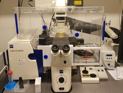
Confocal laser scanning microscope with spectra detector (Meta). Axiovert 200 fully motorized microscope stand, differential interference contrast (DIC), 2 filter based channels with PMT, Meta channel with 32 PMT-array detector. This is an older confocal microscope, but it still performs ok for routine tasks.
Laser lines: 405nm, 458nm, 488nm and 514nm, 543nm and 633nm.
Location: Kunnskapsenter 3rd. floor, room 423.03.031 Confocal Imaging lab
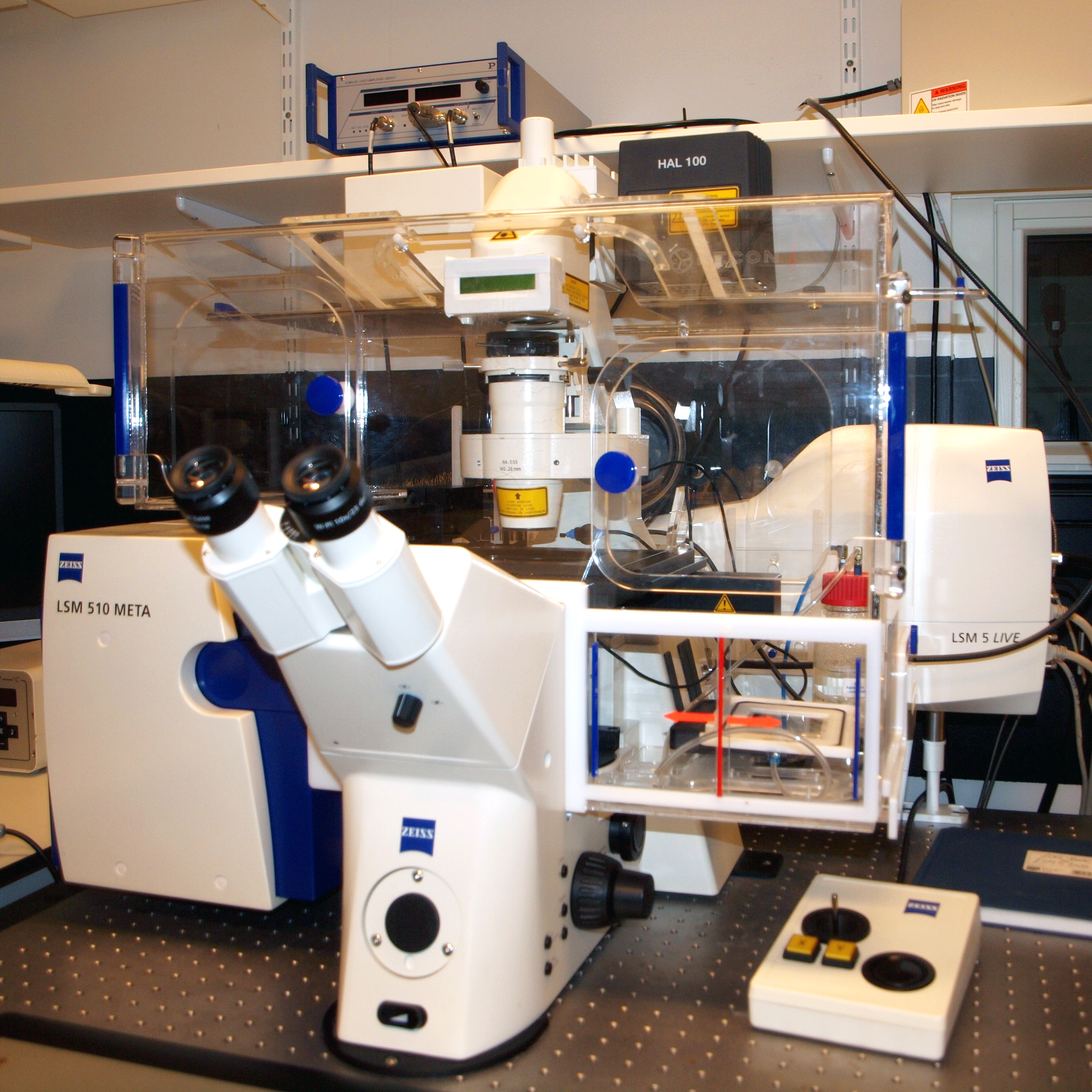 Zeiss 510 META Live
This microscope has not been in active use for a long time, so users need to contact CMIC to perform some tests if they wish to use it.
Confocal laser scanning microscope with spectra detector (Meta) and high speed line scanner (Live). Axiovert 200M fully motorized microscope stand, differential interference contrast (DIC), 2 filter based channels with PMT, Meta channel with 32 PMT-array detector, pinhole in front of all detectors, fast objective piezo z-drive. Laser lines available: Ar 458, 477, 488 and 514nm, diode 561nm and diode 640nm.
Location: Kunnskapssenter 3rd. floor, room 424.03.006A Bio Safety Level 3 lab
This inverted CLSM system is equipped with 3 internal detectors: 2 HyD (high sensitivity, hybrid GaAsP photon counting detectors) and 1 PMTs; high speed resonant scanner (8 KHz) and SuperZ galvo stage for fast lateral and axial acquisition; Adaptive Focus Control (AFC); and a prism-based spectral detection system that enables freely adjustable emission detection bands.
Beam splitters: 15/85, LIAchroic 405/488/552, LIAchroic 405/488/552/638
Objectives: 10x|0.4 (air), 20x|0.75 (multi), 40x|1.3 (oil), 63x|1.4 (oil).
Diode lasers: 405nm (50mW), 488nm (20mW), 552nm (20mW), 638nm (20mW).

Location: Kunnskapsenter 3rd. floor, room 423.03.031 Confocal Imaging lab

This is a microscope with the ability to do image with Total internal reflection fluorescence (TIRF), and is further described under special applications
The Zeiss TIRF 3 is a specialized microscope, but is also well suited for high quality widefield imaging. This is a step up from the simple Evos widefield microscopes, with better light sources, better cameras and increased sensitivity. A black incubator blocks all external light, and allows for stable longterm live imaging under controlled temperature and CO2 conditions.
Objectives: 10x|0.45 (air), 25x|0.8 (multi), 40x|1.4 (oil), 63x|1.3 (oil), 63x|1.46 (oil), 100x|1.57 (oil)
Laser lines available: 405nm (50mW), 488nm (100mW), 561nm (75mW), 638nm (75mW)
Location: Labsenter 4th. floor, room 231.04.037 Histology Lab

This is a widefield (brightfield and 5-channel fluorescence) automated slide scanner tailored for acquiring high volumes of data automatically
General description:
Work in progress
Technical details:
- General:
- Detectors: camera
- Objectives:
- Light source:
Location: Gastro building north, 3rd. floor, St.Olavs hospital
Scan^R offers fully automated high-throughput fluorescence, phase contrast and polarization image acquisition, including time-lapse (in environmental chamber) and automated analysis, so-called high-content screening. Examples of applications: cell transfection efficiency studies, intracellular transport and location studies, RNA interference, bacterial infection, cell cycle.
Location: Gastro building north 3rd. floor, St.Olavs hospital
Inverted fluorescence microscope Olympus IX71, Exfo 120 xenon-Hg excitation light coupled by optic fiber, fast remote controlled excitation filter wheel, mono-, trippel- and quatro- emission filters, polarization filter, phase contrast, objectives with magnification ranging from 4X to 60X, high sensitivity monochrome CCD camera XM10, color CCD camera XC30 and computer with Cell^P Image visualization software.
Location: Kunnskapsenter 3rd. floor, room 423.03.005 Confocal Imaging lab.

Auto Imaging System for simple automated fluorescence imaging in multiwell plates, slides and dishes. Incubator with temperature and CO2 control allows for monitoring of live cells and time-lapse imaging.
The microscope is fully-automated, digital, inverted multi-channel fluorescence and transmitted light imaging system with color and mono cameras. It can do high-resolution mosaic tiling, multi position well scanning, and possibility for cell counting with threshold and multi spot time-lapse studies. The microscope has inserts for slides, petri dishes, 6 – 96 well plates, 25 and 75ml flasks.
8 LED Light cubes with filters for [Ex/Em in nm]: 390/447 (TagBFP), 445/510 (ECFP), 470/510 (EGFP), 500/524 (YFP), 531/593 (RFP), 585/624 (Texas Red), 628/692 (Cy5), 710/775 (Cy7)
Objectives:4x|0.13 (air), 10x|0.25 (air), 20x|0.45 (air), 40x|0.65 (air), 60x|0.75 (air) all with long working distance, no immersion
Location: Labcenter
Auto Imaging System for simple automated fluorescence imaging in multiwell plates, slides and dishes.
This is a simple widefield fluorescence microscope, similar to Evis FL auto 2 but without the incubator option.
The microscope is fully-automated, digital, inverted multi-channel fluorescence and transmitted light imaging system with color and mono cameras. It can do high-resolution mosaic tiling, multi position well scanning, and possibility for cell counting with threshold and multi spot time-lapse studies. The microscope has inserts for slides, petri dishes, 6 – 96 well plates, 25 and 75ml flasks.
8 LED Light cubes with filters for [Ex/Em in nm]: 390/447 (TagBFP), 445/510 (ECFP), 470/510 (EGFP), 500/524 (YFP), 531/593 (RFP), 585/624 (Texas Red), 628/692 (Cy5), 710/775 (Cy7)
Objectives:4x|0.13 (air), 10x|0.25 (air), 20x|0.45 (air), 40x|0.65 (air), all with long working distance, no immersion

Location: Kunnskapssenter 3rd. floor, room 423.03.031 Confocal Imaging lab
Confocal laser scanning microscope equipped with a gated 3D STimulated Emission Depletion (STED) module for super resolution.
The programmable AOBS (acousto-optical beam splitter) provides up to 8 simultaneous excitation lines. The tuneable and pulsed white light laser (WLL) together with the spectral detection system enables freely tunable excitation from 470-670nm (1 nm steps) along with freely adjustable emission detection bands. Additional features include Lightgate functionality, which allows for temporal gating of the excitation light and Lambda Square that enables spectral mapping of excitation and emission information. The system has 4 internal detectors: 2 HyD (high sensitivity, hybrid GaAsP photon counting detectors) and 2 PMTs; high speed resonant scanner (8 KHz) and SuperZ galvo stage for fast lateral and axial acquisition; Adaptive Focus Control (AFC); a Ludin incubation chamber (temperature controlled, active CO2 gas regulator) as well as Huygens deconvolution software for 3D STED data sets.
Objectives: 10x|0.4 (air), 10x|0.4 (multi), 20x|0.75 (multi), 40x|1.3 (oil), 63x|1.4 (oil), 63x|1.3 (glyc), 100x|1.4 (oil).
Laser lines available: 405nm diode, WLL2 (white light laser) tunable from 470-670nm.
STED depletion lasers: 592nm, 660nm, 775nm.
Location: Kunnskapsenter 3rd. floor, room 423.03.031 Confocal Imaging lab
Total internal reflection fluorescence (TIRF) microscope for (long-term) imaging of the plasma membrane region of live cells.
Motorized TIRF slider for reproducible setting of the TIRF angle. This microscope is also equipped with an incubator. Excellent optics for enhanced brightness.Dual cam module with 2x Hamamatsu EMCCD EMX2. DirectFRAP module. Hardware auto-focus Definitive Focus, and software auto-focus
Objectives: 10x|0.45 (air), 25x|0.8 (multi), 40x|1.4 (oil), 63x|1.3 (oil), 63x|1.46 (oil), 100x|1.57 (oil)
TIRF filters: GFP/YFP, CFP/GFP/DsRED, GFP/mRFP/Alexa633, DAPI/FITC/Rhod/CY5
Regular filters:
Laser lines available: 405nm (50mW), 488nm (100mW), 561nm (75mW), 638nm (75mW)

Workstations
Location: Kunnskapsenteret 3rd. floor, room 424.03.024

State-of-the-art workstation aimed at GPU intensive tasks such as deconvolution, denoising and training/fine-tuning deep learning models. Windows 11 OS with WSL2.
Specifications:
- CPU: Intel Xeon w3-2435 8C/16T (3.1GHz Base, 4.5GHz Turbo)
- GPU: Nvidia RTX 6000 Ada 48GB GDDR6 VRAM
- RAM: 128GB DDR5 RAM @ 4800 Mhz
Available software:
- Nikon NIS Elements AR 6.10
- FIJI (ImageJ)
- Mamba-managed Python virtual environments
- VS Code IDE
Location: Gastrosenteret 3rd. floor, room 432.03.014

Workstation aimed at memory and CPU intensive tasks leveraging CPU multithreading and parallelization (i.e. batch image processing via Python scripts). Windows 10 OS.
Specifications:
- CPU: Intel Xeon W-2255 10C/20T (3.7GHz Base, 4.5GHz Turbo)
- GPU: Nvidia RTX 2080Ti 11GB GDDR6 VRAM
- RAM: 128GB DDR5 RAM @ 4800 Mhz
Available software:
- Huygens
- FIJI (ImageJ)
- Cell Profiler
- Microscopy Image Browser
- Cellpose
- Mamba-managed Python virtual environments
- VS Code IDE
Location: Gastrosenteret 3rd. floor, room 432.03.014

Old trusty workstation aimed at 3D image visualization (Imaris) and light image analysis tasks using open source tools. Windows 10 OS.
Specifications:
- CPU: Intel Xeon E5-1660 6C/12T (3.3GHz Base, 3.9GHz Turbo)
- GPU: Nvidia Quadro K5000 4GB GDDR5 VRAM
- RAM: 32GB DDR4 RAM @ 4800 Mhz
Available software:
- Huygens
- Imaris 8.2.1
- ImarisViewer 10.0.1
- LAS X Office
- Cell Profiler
- Dragonfly
- Cellpose
Software
CMIC uses Huygens Professional for deconvolution, image analysis, image processing and visualization.
All our users can get access to Huygens Professional on their own PCs (Windows, Linux or Mac) via the Huygens Everywhere login. Please contact Bjørnar Sporsheim for access and more info about this feature.
Features
- Deconvolution (GPU acceleration, workflow and batch processing, and more)
- 3D visualization (Volume renderer, surface renderer, and more)
- Image Analysis (Interactive object analyzer, colocalization analyzer, object tracker)
- Image processing (bleaching effects, spherical aberration, drift, crosstalk, uneven field illumination, and more)
The Huygens Professional is installed on the Dell Precision 5820 Workstation
For access to Huygens Everywhere please contact Bjørnar Sporsheim.
Book our instruments in
![]()
Contact and Service Request

Manager
Bjørnar Sporsheim
Phone: +47 93033045
Email: bjornar.sporsheim@ntnu.no
CMIC-ALM location
Kunnskapssenteret, 3rd Floor (3. etg.)
St. Olavs Hospital
Olav Kyrres gate 10
7030 Trondheim
Norway
CMIC-Histology location
Laboratoriesenteret, 4th Floor East (4. etg)
St. Olavs Hospital
Erling Skjalgsons gate 1
7030 Trondheim
Norway
Delivery address
NTNU/IKOM
Logistikksenteret Helse Midt Norge
Industriveien 59
Internt: Gastrosenteret 3.etg. sør
7080 Heimdal